77.- HYALINE PLEURAL PLAQUES AND ASBESTOS. REPORT OF TWO FORENSIC CASES. Prof. Garfia.A
77.-PLACAS HIALINAS PLEURALES Y ASBESTOSIS. A PROPÓSITO DE DOS CASOS FORENSES.
Prof. Garfia.A
Prof. Garfia.A
Pneumoconiosis is the deposition of inorganic dust in the lung and the tissue's reaction to its presence. Most dust is deposited in the conducting airways, traped by mucus and removed on the mucociliary defensive system. Soluble dust deposited in alveoli may be cleared by dissolution, but insoluble particles are primarily removed by the alveolar macrophages. The toxicity of dust to the alveolar macrophages varies widely. Most dusts are relatively non-toxic and after phagocitosing the particles the macrophages clears the alveoli by migrating on to the mucociliary system or into the interstitium and after via interstitial lymphatics vessels to the regional lymph nodes. With heavy dust burdens, such as in coal worker's anthracosis, macrophages cannot keep pace with dust intake and particle-laden cells form focal acumulation in the alveoli adjacent to small bronchioles.This dust is relatively non-toxic and cell aggregates (dust cell nests) may remain viable for a considerable time. When cells die they incite little or no fibrosis and remain as non-progressive focal aggregates. However, some dusts, are toxic to alveolar macrophages. The more important group belongs to the crystalline forms of silica, such as quartz, and Asbestos.
Asbestos is a generic name for a group of fibrous silicates. Many of these are fibrogenic, but the relative potency and mechanisms of toxicity are still in some doubt.Although the fibres are cytotoxic to macrophages "in vitro", the correlation with "in vivo" fibrogenicity is low. The physical properties of the fibres appear to be more important and long fibres (> 20um) will be more fibrogenic than shorter ones. These may be too large for complete phagocytosis by the macrophage cells and partly ingested fibres would provide ample opportunity for enzyme leakage and subsequent tissue damage. The tissue reaction in human lungs is variable. The term asbestosis is generally reserved for an interstitial fibrosis which usually begins at the periphery of the lower lobes and progressivelly destroy the respiratory tissue.Other responses, included in the cases studied here, are the existence of hyaline pleural plaques on the parietal pleura and diaphragma, and sometimes the neoformation of a pleural tumor termed "mesothelioma".
 |
| Histopatología Forense Práctica Blog 77. Foto 1 Prof. Garfia.A |
FIG. 1.-
CASE Nº 1.-Male, 20 years old, heroin user and Methadone treatment. The leading cause of death was heroin overdose.
Hyaline pleural plaque located on diaphragmatic surface was an incidental finding of autopsy. It is not know whether there was exposure to asbesto. No "asbestos bodies" were found during microscopic examination of the lungs in this cadaver.
 |
| Histopatología Forense Práctica Blog 77. Foto 2A Prof. Garfia.A |
Fig. 2A.-Case nº 2.- Male, 54 years old.
Representative appearence of hyaline pleural plaques on diaphragmatic surfaces from a man, occupationally exposed to asbestos for many years, worker in naval industry.
The leading cause of death was acute myocardial infarction.
 |
| Histopatología Forense Práctica Blog 77. Foto 2B Prof. Garfia.A |
FIG. 2B.-
Hyaline pleural plaques on diaphragmatic surface -from the same case- showing focal nodular growth. Nodules show morphological aspect of a "nest of round worms".
CROMOPATHOLOGY.
The macroscopical white color is due to the presence of dense fibrous connective tissue plenty of collagen oriented fibers.
Prof.Garfia.A
 |
| Histopatología Forense Práctica Blog 77. Foto 2C Prof. Garfia.A |
Fig. 2C.-Hyaline pleural plaques.
Microscopical section to show the structure of the hyaline plaques and the nodules. Both are composed by dense hypocellular fibrous connective tissue with a basket-weave appearance.
 |
| Histopatología Forense Práctica Blog 77. Foto 2D Prof. Garfia.A |
FIG. 2.D.- Microscopical detail of a fibrous nodule to show that they are composed of dense, relatively hypocellular connective tissue that exhibits basket-weave histological pattern.
Prof. Garfia.A
 |
| Histopatología Forense Práctica Blog 77. Foto 2E Prof. Garfia.A |
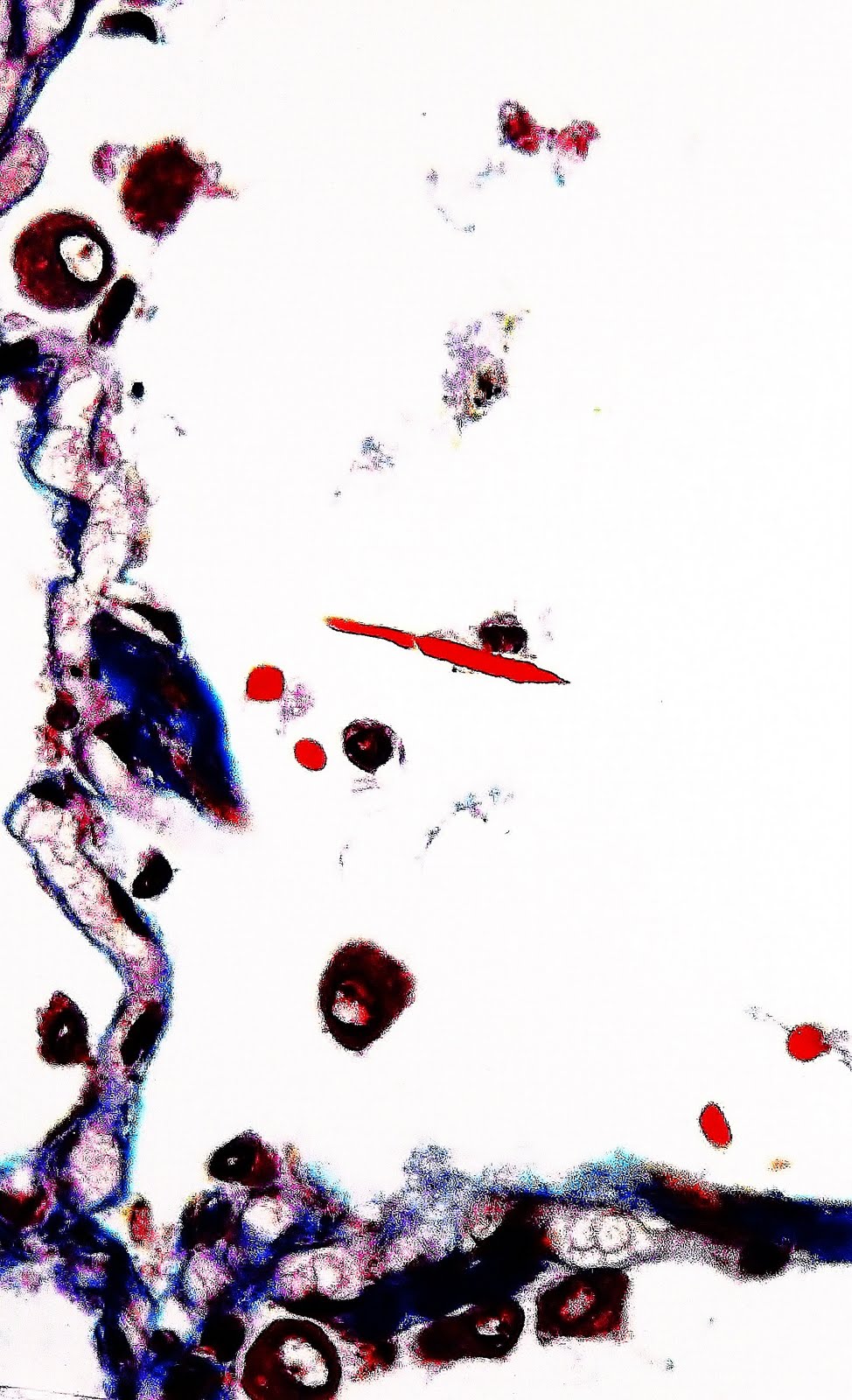
Uncoated fiber, orange-stained, lying free in an alveolar space. Masson Trichrome Stain.
Prof. Garfia.A
 |
| Histopatología Forense Práctica Blog 77. Foto 2E Prof. Garfia.A |
Fig. 2 E.- Lung. Alveolar space.
In this microscopical section we can see another uncoated fiber which is being partially engolfed by a macrophage cell.
Prof. Garfia.A
 |
| Histopatología Forense Práctica Blog 77. Foto 2F Prof. Garfia.A |
Fig. 2F.- Lung. Alveolar space.
Ferruginous body lying free inside an alveolar space. The term "ferruginous body" means a "iron-rich structure". These golden-brown structures were first reported in lung tissue by Marchand in 1906. Cooke, emphasize the relationship of exposure to asbestos dust with the formation of these bodies. These ferruginous structures when seen in tissue sections are routinely classiffied as asbestos bodies. Their presence in tissue is the hallmark of past asbestos exposure. Asbestos is not the only inhaled particulate that stimulates the deposition of a surrounding iron-protein coat. Asbestos bodies are stained with the Perl's method for trivalent iron.
 |
| Histopatología Forense Práctica Blog 77. Foto 2G Prof. Garfia.A |
Fig. 2G.- Lung. Microscopical section.
Ferruginous body showing a typical golden-brown color (CHROMOPATHOLOGY) and a morphological structure which shows striking similarities with a "chess figure".
Prof. Garfia.A
 |
| Histopatología Forense Práctica Blog 77. Foto 2H Prof. Garfia.A |
Fig. 2.H.- Lung. Microscopical section.
Asbestos body partially engolfed by a macrophage cell (arrows).Prof.Garfia.A
 |
| Histopatología Forense Práctica Blog 77. Foto 2I Prof. Garfia.A |
Fig. 2.I.- Lung. Microscopical secion. Masson Trichrome stain.
Asbestos body completelly surrounded by a macrophage cell which contain three or more nuclei (arrows).
Prof.Garfia.A

 |
| Histopatología Forense Práctica Blog 77. Foto 2J Prof. Garfia.A |
Fig. 2.J.- Lung. Microscopical section. Haematoxilin-eosin-phloxine.
To show an asbestos body, phagocited by a macrophage cell, which show a morphological "maraca aspect" and internal structure similar to a truncated pyramid (black and white photo offers the best contrast). (Prof.Garfia.A
 |
| Histopatología Forense Práctica Blog 77. Foto 2K Prof. Garfia.A |
Fig.2K .- Asbestos bodies (ferruginous bodies) and giant cells.
Asbestos bodies are occasionally seen in the cytoplasma of multinucleate histiocytic giant cells often being fragmented as showed in this photograph (arrows).Transverse section of others asbestos bodies are located inside the cytoplasma at three hours of the sphere (star).
Prof. Garfia.A
Prof. Garfia.A
 |
| Histopatología Forense Práctica Blog 77. Foto 2L Prof. Garfia.A |
Fig. 2. L.- Lung. Asbestosis. Microscopical section.
Macrophage cell containing ¿asbestos bodies? inside the lumen of a lymphatic vessel (arrow). Prof. Garfia.A
 |
| Histopatología Forense Práctica Blog 77. Foto 2M Prof. Garfia.A |
Fig. 2.M.- Lung. Asbestosis. Microscopical section.
Vessel containing a ¿giant cell? with the cytoplasm plenty of inclusions pink -stained ¿(asbestos bodies)?
 |
| Histopatología Forense Práctica Blog 77. Foto 2N Prof. Garfia.A |
Fig.2.N.Lung.Asbestosis.Microscopical section.
Vasculitis. Perivascular cuffing (arrows) of lymphocytes, plasma cells and macrophages around the venule (star).
Prof. Garfia.A
Vasculitis. Perivascular cuffing (arrows) of lymphocytes, plasma cells and macrophages around the venule (star).
Prof. Garfia.A
Bibliografía consultada.-
Dail and Hammar's Pulmonary Pathology:Non neoplastic lung disease. Springer 2008. 3ª ed.