29.02.2012
TOXICOLOGICAL PATHOLOGY
118.4.-TOXIC EPIDERMAL NECROLYSIS FATAL AND PHENITOYN.
Prof.Garfia.A
Este caso se publicó -parcialmente- en la versión inglesa del blog "forensicpathologyforum.blogspot.com" con el nº 7. He considerado conveniente traerlo aquí y agruparlo con el resto de casos que forman este grupo de enfermedades raras.
118.4.-NECRÓLISIS EPIDÉRMICA TÓXICA FATAL ASOCIADA A LA ADMINISTRACIÓN DE DILANTIN (FENITOÍNA).
Prof.Garfia.A
TOXICOLOGICAL PATHOLOGY
118.4.-TOXIC EPIDERMAL NECROLYSIS FATAL AND PHENITOYN.
Prof.Garfia.A
CASE REPORT
Untowards reactions to drugs are an important medical problem, sometimes followed by a fatal outcome. Although drugs reactions may involve any organ system, cutaneous eruptions drug-induced may result from a drug administered by any route, which reactions can be identified more frequently than other toxics drug-depending effects which occurs in other organs, such as liver and kidney, because their visibility. Toxic epidermal necrolysis (TEN) or Lyell's Syndrome, is one of the most dramatic, the most severe, and one of most often studied cutaneous drug reactions. TEN is characterized by extensive detachment of the epidermis nuder-going full-thickness necrosis. The drugs more frequently implicated are: anticonvulsivants (phenitoyn, phenobarbital, carbamazepine); antibiotics (ampicillin), sulfonamides and nonsteroids antiinflamatory agents (phenylobutazone, oxyphenbutazone, isoxicam, piroxicam and allopurinol).
I report here a fatal case of TEN related to the anticonvulsivant drug phenitoyn, which was prescribed a man, 74 years old, one month before in order to treat a temporal epilepsy, secondary to a stroke (residual epilepsy).
 |
| PROF. GARFIA. BLOG 118.4 |
Introducción
Las reacciones adversas a los medicamentos constituyen, hoy día, un problema médico sanitario tan grave que puede, a veces, terminar con la vida de un paciente. Es por esta razón por la que el diagnóstico precoz de una tóxicodermia eruptiva fármaco-dependiente es muy importante desde el punto de vista clínico, al objeto de suspender inmediatamente la medicación implicada -y de establecer las medidas terapéuticas correctoras adecuadas- antes de que puedan presentarse complicaciones fatales que podrían desencadenar la puesta en marcha de denuncias judiciales por mala praxis.
Aunque la toxicidad de un fármaco puede afectar a cualesquiera de los sistemas corporales, la "ventaja clínica" que presentan las tóxicodermias eruptivas fármaco-dependientes, que pueden presentarse después de la administración de un fármaco por cualquier vía, es que son visibles. Este hecho permite que se puedan diagnosticar más rápidamente -y más frecuentemente- que las lesiones orgánicas y/o tisulares fármaco-dependientes.
Formas clínicas
Existe una gran variedad de formas clínicas -macroscópicas- de tóxicodermias eruptivas, tantas como erupciones idiopáticas.
Esto significa que una tóxicodermia puede, teóricamente, remedar a cualquier enfermedad eruptiva idiopática de la piel.
Una erupción cutánea puede ser originada por diferentes fármacos (al igual que sucede con las reacciones tisulares y/o orgánicas), y el mismo fármaco puede inducir una gran variedad de patrones eruptivos cutáneo-mucosos. Por estas razones, las tóxicodermias fármaco-dependientes se clasifican, principalmente, de acuerdo con su aspecto macroscópico (clínico), ya que los cambios morfológicos microscópicos son similares, tanto si la enfermedad eruptiva aparece en su forma idiopática como si lo hace después de la administración de una droga -aunque, a veces, el cuadro histopatológico pueda presentar algunas características propias de las reacciones adversas a los medicamentos).
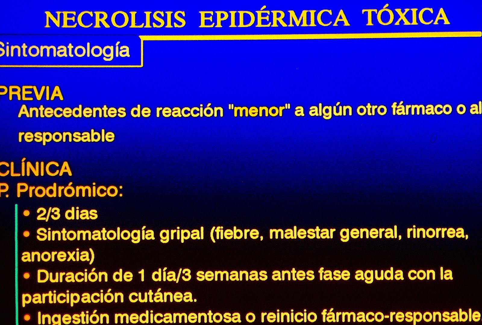
La Necrólisis Epidérmica Tóxica (T.E.N.) o Síndrome de Lyell es la tóxicodermia más dramática, más grave, y una de las tóxicodermias fármaco-dependientes más estudiada.
Las drogas más frecuentemente relacionadas con la aparición de la enfermedad son:
1.-Anticonvulsivantes (fenitoína, fenobarbital, carbamacepina).
2.-Los antibióticos (ampicilinas).
3.-Las sulfonamidas.
4.-Los antiinflamatorios no esteroideos (fenilbutazona, oxifenbutazona, isoxicam, piroxicam y alopurinol).
 |
| PROF. GARFIA.A BLOG 118.4 |
 |
| PROF. GARFIA.A BLOG 118.4 |
 |
| PROF. GARFIA.A BLOG 118.4 |
 |
| PROF. GARFIA.A BLOG 118.4 |
 |
| PROF. GARFIA.A BLOG 118.4 |
 |
| PROF. GARFIA.A BLOG 118.4 |
118.4.-Fig.1.-A.-
To show a diffuse macular and papular erythematous eruption that rapidly becomes confluent and widespread followed by flaccid fluid filled bullae (B) which rapidly ulcerate, leaving painful raw erosions similar to scalding. Mild pressure on erythematous areas may produce (C) detachment of the epidermis (positive Nikolsky sign) and aspect of wet cloth of the epidermis. As result, there may be large areas of exposed dermis resembling a burn (C). Prof.Garfia.A
FIG.2.-
 |
| PROF. GARFIA.A BLOG 118.4 |
The histopathological features of early lesions of toxic epidermal necrolysis are those of an extreme degree of epidermal keratinocyte necrosis (2A), associated with subepidermal blistering (2B, arrows), as a consecuence of basal cell hydropic degeneration.
In more mature lesion, there is complete separation of the epidermis (2C) and the roof of the subepidermal bulla is, usually, necrotic.
2C.-Showing necrotic epidermis completely separated from underlying dermis and to note the paucity of dermal inflamatory infiltrate.
Prof.Garfia.A
In more mature lesion, there is complete separation of the epidermis (2C) and the roof of the subepidermal bulla is, usually, necrotic.
2C.-Showing necrotic epidermis completely separated from underlying dermis and to note the paucity of dermal inflamatory infiltrate.
Prof.Garfia.A
 |
| PROF. GARFIA.A BLOG 118.4 |
 |
| PROF. GARFIA.A BLOG 118.4 |
 |
| PROF. GARFIA.A BLOG 118.4 |
FIG.-----
 |
| PROF. GARFIA.A BLOG 118.4 |
 |
| PROF. GARFIA.A BLOG 118.4 |
FIG.-
 | |
|
 | |
|
 |
| PROF. GARFIA.A BLOG 118.4 |
FIG.-
|
| PROF. GARFIA.A BLOG 118.4 |
FIG.-
 |
| PROF. GARFIA.A BLOG 118.4 |
FIG.-
 |
| PROF. GARFIA.A BLOG 118.4 |
 |
| BLOG 118.4 PROF. GARFIA.A |
 |
| BLOG 118.4 PROF. GARFIA. |
FIG.- DIAGNÓSTICO DIFERENCIAL
 |
| PROF. GARFIA.A BLOG 118.4 |
FIG.-